
Wound Dressings: Hydrocolloid

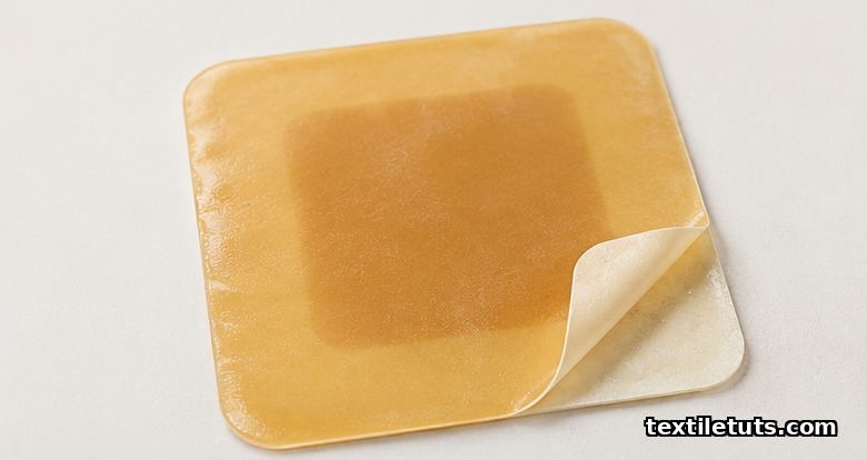
A hydrocolloid wound dressing is a moisture-retentive, semi-occlusive laminate that pairs an elastomer adhesive matrix with embedded hydrophilic colloid particles (sodium carboxymethylcellulose, gelatin, and pectin) and absorbs 10 to 30 times its own weight in wound exudate while sustaining a moist interface with a measured moisture vapor transmission rate of 200 to 700 grams per square meter per 24 hours.
On contact with wound fluid the dry particles swell and fuse into a soft, non-adherent gel that holds the wound bed at a near-neutral pH of 6.1 to 7.5 and a stable local temperature of 32 to 34 degrees Celsius, allowing wear times of 3 to 7 days between dressing changes.
This guide walks textile and apparel students, material scientists, and medical-textile engineers through the layered construction, polymer chemistry, performance testing, and standards that define hydrocolloid dressings as a category of advanced wound care.
What Is a Hydrocolloid Wound Dressing?
Hydrocolloid dressings are a class of advanced wound care product classified under medical textiles by ISO 13485 (quality management for medical devices) and regulated in the United States under FDA 21 CFR 880.5240. They sit alongside alginate, foam, hydrogel, and transparent film dressings but are distinguished by their use of dry, hydrophilic colloidal particles that absorb exudate and convert it into a gel.
The first commercial hydrocolloid was introduced in the early 1980s under the DuoDERM brand (ConvaTec). Within a decade, hydrocolloid technology had become a standard option for low-to-moderately exuding wounds, partial-thickness burns, and pressure ulcer prophylaxis. The category remains one of the most studied sub-segments in advanced wound care literature.
Material Composition and Layered Structure
A hydrocolloid dressing is a multi-layer laminate, typically 0.5 mm to 1.5 mm in total thickness, where every layer serves a defined engineering function:
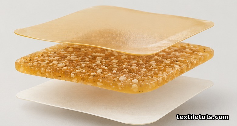
- Outer backing film: a continuous polyurethane (PU) or polyethylene film, 25 to 50 micrometers thick, providing a bacterial and water barrier while remaining vapor-permeable.
- Hydrocolloid adhesive layer: a pressure-sensitive elastomer matrix (commonly styrene-isoprene-styrene block copolymer, or SIS) loaded with 30 to 60 percent by weight of hydrophilic colloid particles.
- Hydrocolloid particles: sodium carboxymethylcellulose (CMC), gelatin, pectin, and optionally guar gum, with particle sizes typically between 10 and 100 micrometers.
- Release liner: a silicone-coated paper or polymer film that protects the adhesive before use and is removed at the point of application.
The hydrocolloid-to-elastomer ratio is a tightly controlled parameter. Higher colloid loadings (above 50 percent) raise absorbency but reduce cohesive strength and increase residue left on the wound bed at dressing change. Lower loadings give cleaner removal at the cost of total fluid handling capacity.
The Absorption and Gelation Mechanism
When wound exudate enters the dressing, water is taken up by the hydrocolloid particles through a combination of capillary action and osmotic swelling. Sodium carboxymethylcellulose alone absorbs 8 to 12 g water per gram of dry polymer, while gelatin absorbs 5 to 10 g/g. The blended formulation typically reaches 10 to 30 g/g overall, depending on particle size distribution and elastomer matrix density.
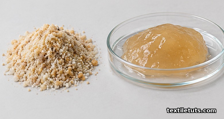
As the particles swell they lose their discrete identity and fuse into a continuous soft gel that conforms to the wound surface. This gel layer performs three clinically important functions:
- Maintains a moist environment that supports autolytic debridement of necrotic tissue.
- Provides thermal insulation, stabilising the wound bed at 32 to 34 degrees Celsius against a core body temperature of 37 degrees Celsius.
- Holds a slightly acidic to neutral pH of 6.1 to 7.5, which has been shown to suppress bacterial proliferation relative to the alkaline environment that develops under dry gauze.
Because the gel is non-adherent to granulation tissue, removal typically does not disturb the healing bed, which is the principal advantage of hydrocolloid technology over traditional woven cotton gauze.
Key Performance Properties
Hydrocolloid dressings are specified by a measurable set of textile and material parameters:
- Moisture vapor transmission rate (MVTR): 200 to 700 g/m squared per 24 h, measured per ASTM E96 (water method) or the equivalent ISO 2528.
- Absorbency: 10 to 30 g of saline per 100 cm squared of dressing over 24 hours under standard test conditions.
- Tensile strength of the backing film: 30 to 60 N per cm of width, measured per ASTM D882.
- Peel adhesion to skin: 0.5 to 4 N per 25 mm, tuned to remain secure during wear but release without trauma on removal.
- Bacterial barrier performance: outer film passes ASTM F903 (liquid penetration) and ASTM F1671 (viral penetration using a bacteriophage model) when tested as a protective material.
- Biocompatibility: cytotoxicity, sensitisation, and irritation tested per ISO 10993-5, ISO 10993-10, and ISO 10993-23.
Manufacturing Process
Industrial production of hydrocolloid dressings follows a continuous, roll-to-roll lamination sequence:
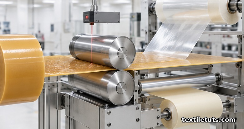
- Hydrocolloid compounding: CMC, gelatin, and pectin are blended with the elastomer adhesive in a sigma-blade or planetary mixer under controlled humidity (below 30 percent RH) to prevent premature moisture pickup.
- Mass extrusion and calendering: the compounded mass is extruded or calendered into a uniform sheet, typically 0.4 to 1.2 mm thick, with online gauge monitoring.
- Laminating with the backing film: the sheet is laminated to a polyurethane or polyethylene film using a heated nip roll at 80 to 120 degrees Celsius.
- Die cutting and release liner application: the laminate is cut to shape (square, sacral, oval, or anatomical) and a silicone release liner is applied.
- Packaging: individual pouches are sealed at low humidity, typically below 20 percent RH, to preserve shelf-life absorbency.
Process control focuses on relative humidity, mass uniformity, and peel adhesion. Inline near-infrared sensors measure water content of the compounded mass in real time, and laser gauges verify calendered thickness within plus or minus 0.05 mm.
Comparison: Hydrocolloid vs. Other Advanced Dressings
| Property | Hydrocolloid | Alginate | Foam (PU) | Hydrogel |
|---|---|---|---|---|
| Absorbency (g per 100 cm squared per 24 h) | 10 to 30 | 15 to 40 | 20 to 50 | 5 to 15 |
| MVTR (g/m squared per 24 h) | 200 to 700 | 800 to 1500 | 1500 to 3000 | 2500 to 3500 |
| Wear time (days) | 3 to 7 | 1 to 3 | 3 to 7 | 1 to 3 |
| Best-fit wound type | Low to moderate exudate | Moderate to heavy exudate | Moderate to heavy exudate | Dry or lightly exuding |
| Forms gel on contact | Yes | Yes | No | Already a gel |
| Adhesion | Self-adherent | Needs secondary fixation | Border variants self-adhere | Needs secondary fixation |
Standards That Apply
Hydrocolloid dressings are medical devices, so they are subject to a layered testing regime that pulls from textile, material, and biological evaluation standards:
- ISO 10993 series: biological evaluation of medical devices, including cytotoxicity (Part 5), sensitisation (Part 10), and irritation (Part 23).
- ISO 13485: quality management systems for medical device manufacturers.
- ASTM E96: gravimetric determination of water vapor transmission rate, the basis for MVTR reporting on backing films.
- ASTM F903: resistance of materials used in protective clothing to penetration by liquids.
- ASTM F1671: resistance of materials to penetration by blood-borne pathogens using a bacteriophage model.
- USP <151>: pyrogen test where the dressing is labelled non-pyrogenic.
Frequently Asked Questions
What is the difference between a hydrocolloid and a hydrogel dressing?
Hydrocolloid dressings start as a dry adhesive sheet containing absorbent particles that swell into a gel only after contact with wound fluid. Hydrogel dressings are already 70 to 90 percent water in their supplied state and donate moisture to the wound rather than absorbing it, which makes hydrogels the better choice for dry, necrotic, or lightly exuding wounds.
How long can a hydrocolloid dressing stay on?
Most hydrocolloid dressings are approved for wear times of 3 to 7 days, depending on exudate level, anatomical location, and the manufacturer’s instructions for use. The most common change triggers are visible saturation reaching within 1 cm of the dressing edge, gel pooling, or loosening of the adhesive border.
Can hydrocolloid dressings be used on infected wounds?
Because hydrocolloid dressings create a semi-occlusive environment, they are not recommended for clinically infected wounds without concurrent systemic antibiotic therapy. Consensus guidance from the Wound, Ostomy and Continence Nurses Society (WOCN) advises against using occlusive dressings as a standalone on heavily infected or heavily exuding wounds.
Do hydrocolloid dressings leave residue in the wound?
Older formulations with high gelatin content were known to leave visible gel residue that was sometimes mistaken for slough. Modern carboxymethylcellulose-dominant blends with cohesive elastomer matrices leave minimal residue, and any small amount of remaining gel is non-adherent and rinses away with normal saline irrigation.
Are hydrocolloid dressings breathable?
They are semi-permeable rather than fully breathable. The backing film allows moisture vapor transmission of 200 to 700 g/m squared per 24 h, which is enough to prevent peri-wound maceration but well below the transmission rate of intact skin, measured at roughly 500 to 2000 g/m squared per 24 h depending on body site.
References
- ASTM International. ASTM E96 / E96M-23: Standard Test Methods for Gravimetric Determination of Water Vapor Transmission Rate of Materials. ASTM International, West Conshohocken, PA, 2023.
- ASTM International. ASTM F903-18: Standard Test Method for Resistance of Materials Used in Protective Clothing to Penetration by Liquids. ASTM International, West Conshohocken, PA, 2018.
- ASTM International. ASTM F1671 / F1671M-22: Standard Test Method for Resistance of Materials Used in Protective Clothing to Penetration by Blood-Borne Pathogens Using a Bacteriophage Penetration Model. ASTM International, West Conshohocken, PA, 2022.
- International Organization for Standardization. ISO 10993-5:2009, Biological Evaluation of Medical Devices, Part 5: Tests for In Vitro Cytotoxicity. ISO, Geneva, 2009.
- International Organization for Standardization. ISO 13485:2016, Medical Devices, Quality Management Systems, Requirements for Regulatory Purposes. ISO, Geneva, 2016.
- Thomas S. Hydrocolloid dressings in the management of acute and chronic wounds. British Journal of Nursing. 2020;29(15):S28-S34.
- Queen D, et al. pH changes in wounds after the application of hydrocolloid dressings. Journal of Wound Care. 2019;28(5):316-321.
This article is the working reference for hydrocolloid wound dressings as a category of medical textile. Editorial by Md. Asad Ullah (Meem), TextileTuts. Sources: ASTM E96, ASTM F903, ASTM F1671, ISO 10993-5, ISO 13485, and the cited peer-reviewed wound-care literature as referenced above.


