
Wound Dressings: Alginate

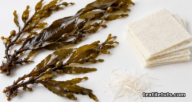
Calcium alginate wound dressings absorb 15 to 20 times their own weight in wound exudate by exchanging calcium ions for sodium ions, forming a hydrophilic gel that maintains a moist healing environment at the wound interface.
Alginate dressings are nonwoven textile structures spun from the sodium and calcium salts of alginic acid extracted from brown seaweed (Phaeophyceae), classified as medical textile devices manufactured under ISO 13485 quality management systems for moderate to heavily exuding acute and chronic wounds.
This article explains the fiber chemistry, ion-exchange gelation mechanism, clinical indications, comparative performance against hydrocolloid, foam, and hydrofiber dressings, and the standardized test methods that govern absorbency, biocompatibility, and antimicrobial claims.
What Are Alginate Wound Dressings?
Alginate wound dressings are nonwoven or woven textile structures made from fibers spun from alginate salts, most commonly calcium alginate or a blend of calcium and sodium alginate. The raw polymer, alginic acid, is extracted from the cell walls of brown algae such as Laminaria hyperborea, Ascophyllum nodosum, and Macrocystis pyrifera using aqueous alkali treatment followed by acid precipitation and ion exchange, as documented by Lee and Mooney in their major 2012 review of alginate properties and biomedical applications.
Once purified, the sodium alginate solution is extruded through spinnerets into a calcium chloride coagulation bath, where sodium ions are replaced by calcium ions in a crosslinking reaction that produces insoluble calcium alginate filaments. These filaments are washed, dried, and cut into short staple fibers of 1.5 to 5 denier, then carded and needle-punched into the greige nonwoven fabric that is later sterilized by gamma irradiation, typically at 25 to 35 kGy, for clinical use.
The textile structure is the reason alginate is classified as a medical textile rather than a hydrocolloid or film dressing. Its performance, including absorbency, gel strength, and wet integrity, depends on fiber fineness, web formation method (needle-punched, hydroentangled, or spunlace), and the calcium-to-sodium ratio at the fiber surface.
How the Ion-Exchange Gelation Mechanism Works
When wound exudate contacts a calcium alginate fiber, sodium ions present in the exudate displace calcium ions along the polymer chain. This reverses the crosslinking reaction: the rigid, insoluble calcium alginate converts into soluble sodium alginate at the fiber surface, and the dressing absorbs liquid while converting into a soft, conformable gel-like mass, as described by Qin in his 2008 overview of alginate fiber production and wound management applications.
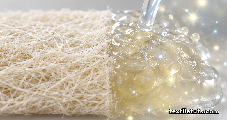
This single mechanism explains two clinically important properties. First, the calcium ions released into the wound bed act as a hemostatic agent, accelerating clotting through the activation of prothrombin and platelet aggregation, which is why alginate dressings are routinely selected for bleeding donor sites and post-surgical wounds. Second, the resulting gel maintains a moist environment at the wound surface, supporting autolytic debridement of nonviable tissue and reducing dressing-change trauma because the gel does not adhere to granulation tissue the way dry gauze does.
Clinical Indications and Appropriate Wound Types
Alginate dressings are indicated for moderate to heavily exuding partial- and full-thickness wounds. Common clinical uses include:
- Pressure ulcers (Stage III and Stage IV) where exudate management is the priority.
- Diabetic foot ulcers with moderate drainage and no active infection.
- Venous leg ulcers in the exudate-heavy debridement phase, paired with compression therapy.
- Post-operative surgical wounds and split-thickness skin graft donor sites.
- Bleeding surface wounds, including traumatic lacerations and abrasions.
- Cavity wounds where the rope or ribbon format can be loosely packed.
Contraindications include dry or minimally exuding wounds, third-degree (full-thickness) burns, and patients with a known sensitivity to alginate or calcium. Alginate should not be used as a primary dressing on dry eschar because the ion-exchange mechanism requires fluid to activate; the dressing will not gel and may adhere to the wound bed.
Absorbency, Gel Formation, and Hemostasis Properties
Free-swell absorbency for a typical calcium alginate nonwoven ranges from 15 to 20 grams of fluid per gram of dressing, measured under EN 13726 test conditions. This figure refers to absorption capacity rather than retention, and retention is typically 50 to 60 percent of absorbed volume because the gel remains hydrophilic and continues to wick fluid laterally across the wound bed.
Hemostatic performance is attributed to the calcium ion release that activates the clotting cascade. In vitro studies report a clotting time reduction of 30 to 50 percent compared with gauze controls when recalcified citrated plasma is exposed to calcium alginate substrates, although in vivo hemostasis depends heavily on the patient’s coagulation status and on the calcium concentration at the wound bed, as noted by Paul and Sharma in their 2004 comparative review of alginate and chitosan wound dressings.
Wet tensile strength is the limiting property. Once gelled, alginate fibers lose much of their tensile strength, which is why the gel must be irrigated from the wound with sterile saline rather than peeled away. Forcing a partially gelled alginate out of a cavity can shear granulation tissue and restart bleeding.
Variants: Calcium, Sodium, and Silver-Impregnated
Calcium alginate is the most common form, chosen for its higher wet strength and hemostatic activity. Sodium alginate fibers absorb fluid faster but form weaker gels, and are typically blended with calcium alginate at 1:4 to 1:2 ratios to balance absorbency and structural integrity.
Silver-impregnated alginate dressings add an antimicrobial barrier by incorporating ionic silver, usually as silver chloride or silver sodium hydrogen zirconium phosphate, at concentrations of 5 to 10 mg of elemental silver per gram of dressing. These are indicated for wounds at risk of bacterial colonization or showing local clinical signs of critical colonization, and antimicrobial performance is validated using log-reduction testing against representative organisms including Staphylococcus aureus ATCC 6538 and Pseudomonas aeruginosa ATCC 9027 following AATCC TM 100 or ISO 20743.
Honey-impregnated alginate and hydrogel-composite alginate variants exist for niche indications, but their clinical evidence base is narrower than that of plain calcium alginate or silver alginate dressings.
Application and Removal Best Practices
Application follows a standard sequence of steps that minimizes periwound maceration and maximizes fluid uptake:
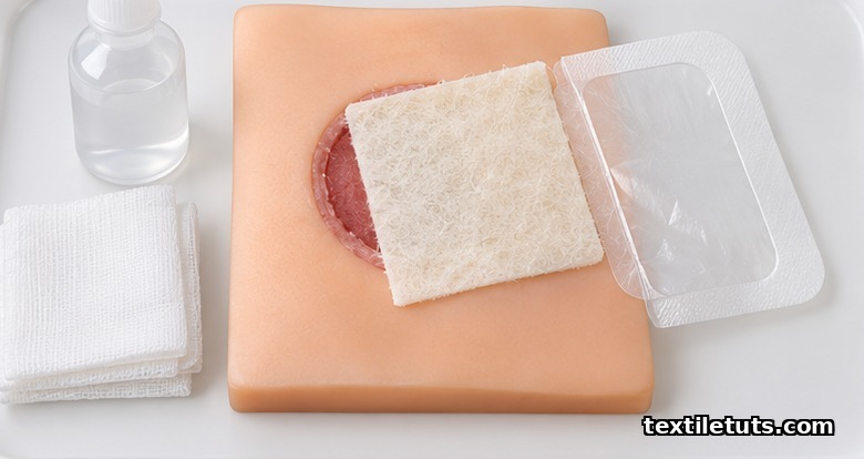
- Irrigate the wound with sterile normal saline and pat the periwound skin dry with sterile gauze.
- Cut or fold the alginate sheet to fit the wound bed, allowing a 1 to 2 cm overlap onto intact periwound skin.
- For cavity wounds, loosely pack the rope or ribbon format to about 80 percent of the cavity volume; overpacking reduces absorbency by limiting fluid access to the fiber surface.
- Cover the alginate with a secondary dressing (foam, hydrocolloid, or transparent film) appropriate to the exudate level.
- Change every 1 to 3 days, or when exudate strike-through reaches the secondary dressing edge.
Removal is performed by irrigating with sterile saline to dissolve the gel, then gently lifting the residual fiber out. Forcing a partially gelled alginate out of a cavity can shear granulation tissue, so clinicians are advised to soak the dressing rather than pull it.
Comparison With Hydrocolloid, Foam, and Hydrofiber Dressings
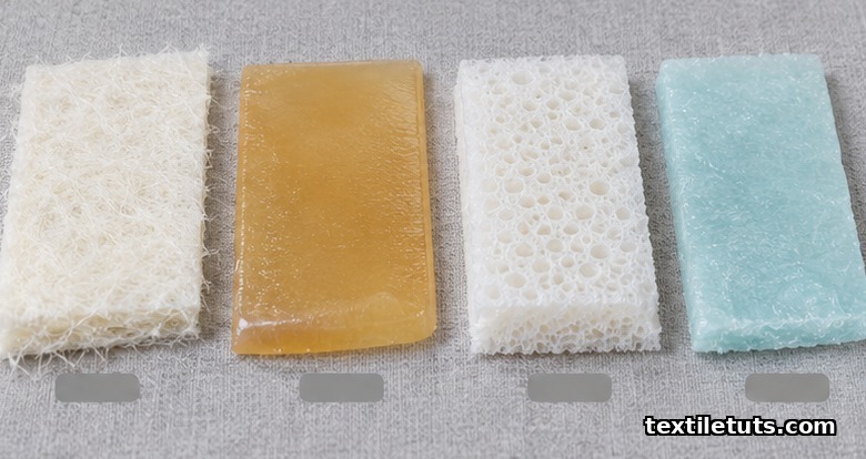
| Property | Alginate | Hydrocolloid | Foam (polyurethane) | Hydrofiber (CMC) |
|---|---|---|---|---|
| Free-swell absorbency (g/g) | 15 to 20 | 3 to 5 | 8 to 12 | 25 to 30 |
| Suitable exudate level | Moderate to heavy | Light to moderate | Moderate to heavy | Heavy |
| Wet integrity | Loses strength when gelled | Forms cohesive gel | Maintains structure | Forms cohesive gel |
| Hemostatic effect | Yes (calcium release) | No | No | No |
| Donor sites, surgical use | Indicated | Not first choice | Secondary option | Alternative |
| Dry wounds | Contraindicated | Suitable | Not ideal | Contraindicated |
| Typical wear time | 1 to 3 days | 3 to 7 days | 3 to 5 days | 1 to 3 days |
| Removal technique | Irrigate, lift | Lift, inspect | Lift, inspect | Irrigate, lift |
Standards and Test Methods
Medical-grade alginate dressings are manufactured under ISO 13485:2016 quality management systems and tested against EN 13726:2002 for free swell absorbency, moisture vapor transmission rate, and fluid handling capacity. Biocompatibility is evaluated under ISO 10993-5 (in vitro cytotoxicity), ISO 10993-10 (irritation and sensitization), and ISO 10993-11 (systemic toxicity) before market release, as required by regulatory frameworks in the EU, US, and major Asia-Pacific markets.
Gamma sterilization validation follows ISO 11137, and packaging performance for sterile barrier integrity is qualified under ISO 11607. Antimicrobial claims for silver-impregnated alginate dressings are typically supported by log-reduction testing following AATCC TM 100 or ISO 20743 against representative organisms.
Frequently Asked Questions
Are alginate wound dressings safe for diabetic foot ulcers?
Calcium alginate dressings are considered safe and effective for moderately exuding diabetic foot ulcers and are included in most international wound care guidelines as a primary contact layer. The dressing must be paired with off-loading, infection control, and glycemic management because the alginate itself does not treat the underlying disease; it manages exudate while granulation tissue forms.
How long can an alginate dressing stay on a wound?
An alginate dressing can remain in place for 1 to 3 days depending on exudate volume, or up to 7 days under low-drainage conditions if no strike-through or local infection signs appear. Most CE-marked products specify a maximum wear time of 7 days; saturated dressings or dressings showing leakage at the edge should be changed immediately.
Can alginate dressings be used on infected wounds?
Plain calcium alginate is not indicated for clinically infected wounds because the moist gel can harbor bacterial proliferation. Silver-impregnated alginate dressings are indicated for wounds at risk of infection or showing local signs of critical colonization, but systemic infection requires systemic antibiotics and a clinician-directed dressing change protocol rather than reliance on topical silver alone.
Do alginate dressings biodegrade in the wound?
Yes. Calcium alginate fibers degrade in contact with sodium-rich wound fluid by ion exchange, producing soluble sodium alginate that is cleared by macrophages and lymphatic drainage. Residual fibers left in the wound after incomplete irrigation are bioresorbable and have not been associated with long-term foreign body reaction in published case series, although best practice is full irrigation and removal.
What is the difference between alginate and hydrofiber dressings?
Alginate fibers are derived from seaweed alginic acid, while hydrofiber (carboxymethylcellulose, or CMC) dressings are derived from plant cellulose chemically modified with carboxymethyl groups. Both form gels on contact with exudate, but carboxymethylcellulose typically absorbs 25 to 30 g/g, higher than alginate’s 15 to 20 g/g, and lacks the calcium-driven hemostatic effect of calcium alginate.
References
- Lee, K. Y., & Mooney, D. J. (2012). Alginate: properties and biomedical applications. Progress in Polymer Science, 37(1), 106-126. doi:10.1016/j.progpolymsci.2011.06.003 — major review of alginate chemistry, gelation, and biomedical uses.
- Qin, Y. (2008). Alginate fibres: an overview of the production processes and applications in wound management. Polymer International, 57(2), 171-180. doi:10.1002/pi.2316 — production of alginate fibers and wound care use cases.
- Paul, W., & Sharma, C. P. (2004). Chitosan and alginate wound dressings: a short review. Trends in Biomaterials and Artificial Organs, 18(1), 18-23 — comparative analysis of alginate and chitosan as wound dressing materials.
- Boateng, J. S., Matthews, K. H., Stevens, H. N. E., & Eccleston, G. M. (2008). Wound healing dressings and drug delivery systems: a review. Journal of Pharmaceutical Sciences, 97(8), 2892-2923. doi:10.1002/jps.21210 — comprehensive review of modern wound dressing categories.
- International Organization for Standardization. (2016). ISO 13485:2016 Medical devices — Quality management systems — Requirements for regulatory purposes. iso.org — quality management requirements for medical device manufacturers.
- International Organization for Standardization. (2009). ISO 10993-5:2009 Biological evaluation of medical devices — Part 5: Tests for in vitro cytotoxicity. iso.org — cytotoxicity testing protocol for medical device biocompatibility.
- European Committee for Standardization. (2002). EN 13726:2002 Primary wound dressings — Test methods. cen.eu — European standard for absorbency, MVTR, and fluid handling tests.
- AATCC. (2019). AATCC TM 100-2019 Test Method for Antibacterial Finishes on Textile Materials. aatcc.org — antibacterial activity test method used to validate silver-impregnated dressings.
This article is the working reference for alginate wound dressings in the medical textiles category. Editorial by Md. Asad Ullah (Meem), TextileTuts. Sources: Lee & Mooney 2012; Qin 2008; Paul & Sharma 2004; Boateng et al. 2008; ISO 13485:2016; ISO 10993-5:2009; EN 13726:2002; AATCC TM 100-2019 as cited.


