
Tissue Engineering Scaffolds

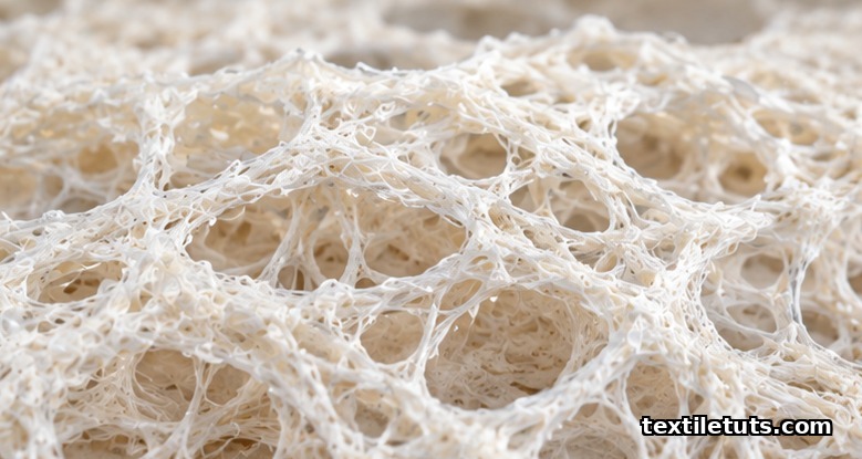
Tissue engineering scaffolds are three-dimensional porous structures, typically fabricated with fiber diameters from 50 nm to 500 μm and pore sizes of 50–500 μm, that support cell attachment, proliferation, and guided regeneration of skin, bone, cartilage, vascular, and nerve tissue.
Textile manufacturing methods such as electrospinning, weaving, knitting, and braiding are the primary routes to scaffold fabrication because they deliver precise control over fiber architecture, mechanical anisotropy, and degradation kinetics using biocompatible polymers such as polycaprolactone (PCL), poly(lactic-co-glycolic acid) (PLGA), and collagen.
This article covers the textile-engineering perspective on scaffold design: how yarn and nonwoven formation methods shape scaffold properties, why pore size and porosity determine tissue outcome, the ASTM and ISO standards that govern testing, and the material choices that match specific clinical targets.
What a Tissue Engineering Scaffold Is
A scaffold is a temporary, biodegradable matrix that mimics the extracellular matrix (ECM) of the target tissue. Its three jobs are to provide mechanical support during healing, to deliver cells and signaling molecules to the defect site, and to degrade at a rate matched to new tissue deposition. The global market for tissue-engineered products reached roughly USD 12.5 billion in 2024, with scaffold-based products representing the largest commercial segment.
Scaffolds sit at the intersection of textiles, materials science, and cell biology. Because they are implantable, they must pass biological evaluation under ISO 10993 and meet characterization requirements described in ASTM F2150. The geometry is not aesthetic; every fiber diameter, pore size, and degradation rate ties directly to a clinical outcome such as healing time, mechanical load-bearing, or graft integration with host tissue.
Textile Manufacturing Methods for Scaffolds
Four textile routes dominate scaffold fabrication, each producing a different fiber architecture.
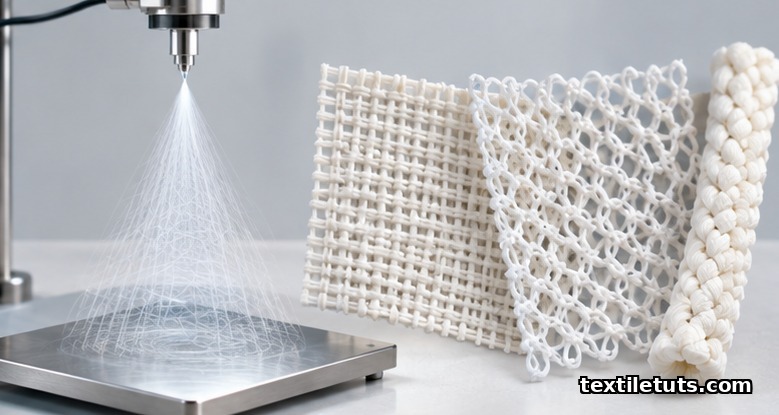
Electrospinning produces a nonwoven mesh of continuous fibers with diameters typically between 50 nm and 1 μm. The process uses an electric field of 10–30 kV to draw a polymer solution from a spinneret onto a grounded collector, generating mats with porosity above 80% and specific surface areas of 10–100 m²/g. The nano-scale fiber diameter mimics native collagen fibrils (50–500 nm), which is why electrospun mats are favored for skin, nerve, and cardiac patches.
Weaving uses two orthogonal yarn systems (warp and weft) to build dense, mechanically robust scaffolds with tensile strengths up to 50 MPa for PLGA yarns. Woven scaffolds carry load well but have lower porosity (typically 30–60%), so they are reserved for load-bearing tissues such as tendon, ligament, and bone.
Knitting interlopes yarns in a loop structure, delivering high porosity (60–85%) and elasticity. Knitted vascular grafts and hernia meshes are commercial products because the loop architecture permits suture retention and burst-pressure resistance above 200 kPa.
Braiding twists three or more yarn sets to make tubular scaffolds with controllable radial compliance, used primarily for tendon and ligament reconstruction.
Other methods include 3D printing (extrusion of polymer melt or solution layer by layer) and freeze-drying (ice-templating of a polymer slurry to leave a porous foam). Hybrid scaffolds combine two or more of these routes; for example, a knitted PCL tube wrapped in an electrospun collagen mat to combine mechanical compliance with cell-recognition signaling.
Material Selection: Natural vs Synthetic Polymers
Material choice controls degradation rate, mechanical stiffness, and cell-recognition signals.
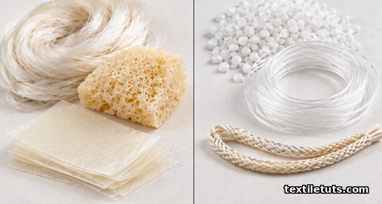
Natural polymers such as collagen, chitosan, gelatin, silk fibroin, and hyaluronic acid carry built-in cell-binding motifs (for example, RGD on collagen) and degrade by enzymatic hydrolysis. Collagen sponges, derived from bovine or porcine sources, have been used in skin and cartilage repair for decades. Silk fibroin, degummed from Bombyx mori cocoons, has tensile strength up to 500 MPa, making it the strongest natural scaffold fiber.
Synthetic polymers such as polycaprolactone (PCL), polylactic acid (PLA), polyglycolic acid (PGA), and PLGA offer tunable degradation (from months to years via copolymer ratio), reproducible batch quality, and mechanical properties that can be matched to host tissue. PCL degrades in roughly 2–4 years, PLA in 12–24 months, PGA in 2–4 months, and PLGA tunable across that window.
Composite scaffolds combine a synthetic framework with a natural coating. A PCL nanofiber mat coated with gelatin, for example, gains the RGD cell-binding sites of gelatin while keeping the mechanical strength of PCL. Composite designs are now standard in research publications and emerging commercial products.
Key Scaffold Properties: Porosity, Pore Size, Mechanical Match
Three measurable properties drive clinical performance.
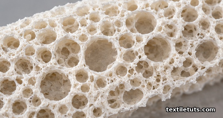
Porosity describes the void fraction of the scaffold; values of 60–90% are typical. Higher porosity improves mass transport and cell migration but reduces mechanical stiffness, so design selects the minimum porosity that still supports nutrient diffusion throughout the implant.
Pore size is more critical than porosity alone. Bone regeneration needs pores of 200–500 μm to allow osteoblast infiltration and vascular ingrowth. Cartilage regenerates in pores of 100–250 μm. Skin regeneration works with pores of 20–125 μm. Nerve guidance channels use aligned microchannels of 5–30 μm diameter to direct axon growth along the channel axis.
Mechanical strength must match host tissue to avoid stress shielding. Scaffolds for articular cartilage target a compressive modulus near 0.5–1 MPa. Cancellous bone scaffolds target 50–500 MPa. Suture retention strength above 2 N is required for any load-bearing scaffold, per ASTM F2150. The mismatch in stiffness between scaffold and host tissue drives scar formation rather than regeneration.
ASTM and ISO Standards for Scaffold Testing
Regulatory bodies require standardized characterization before any implant can be studied in humans. Three standards form the baseline.
ASTM F2150 (“Standard Guide for Characterization and Testing of Biomaterial Scaffolds Used in Tissue-Engineered Medical Products”) defines the minimum physical, mechanical, and biological characterization. It requires reporting of material composition, scaffold architecture (fiber diameter, pore size, porosity), degradation profile, and cytocompatibility per ISO 10993.
ASTM F2739 (“Standard Guide for Quantitating Cell Viability Within Biomaterial Scaffolds”) prescribes live/dead cell assays, MTT, and confocal imaging protocols to measure cell distribution and viability across the full 3D scaffold thickness, not just the surface layer.
ISO 10993 (“Biological evaluation of medical devices”) sets the toxicity, sensitization, irritation, genotoxicity, and implantation-testing framework that all medical textile scaffolds must pass before clinical use. ISO 10993-1:2018 is the current version of the evaluation-and-testing guideline.
Comparison: Textile Scaffold Fabrication Methods
| Method | Fiber / Yarn Geometry | Typical Fiber Diameter | Porosity | Mechanical Strength | Best-Fit Tissue |
|---|---|---|---|---|---|
| Electrospinning | Nonwoven nanofiber mat | 50 nm – 1 μm | 80–95% | Low tensile, high surface area | Skin, nerve, cardiac patch |
| Weaving | Warp + weft orthogonal | 10 – 500 μm | 30–60% | High (up to 50 MPa) | Tendon, ligament, bone |
| Knitting | Interlooped yarns | 10 – 500 μm | 60–85% | High compliance | Vascular graft, hernia mesh |
| Braiding | Twisted tubular | 10 – 500 μm | 40–70% | Tuned radial stiffness | Tendon, ligament |
| 3D printing | Layer-by-layer extrusion | 100 – 500 μm | 50–80% | Tunable by infill | Bone, cartilage, complex 3D shapes |
| Freeze-drying | Foam / ice-templated | Continuous foam | 70–95% | Low (brittle) | Soft tissue, drug delivery |
Frequently Asked Questions
How long does a tissue engineering scaffold last inside the body?
Degradation time is set by polymer choice and scaffold architecture. PGA scaffolds lose 50% of their mass in 2–4 weeks; PLA scaffolds in 6–12 months; PLGA between those windows; PCL in 2–4 years. The design target is for the scaffold to degrade at a rate matched to new tissue deposition, so mechanical load transfers gradually to the regenerating tissue rather than collapsing into a void.
Are tissue engineering scaffolds approved for clinical use?
Yes. Approved products include Apligraf (a bilayered skin substitute using a bovine collagen matrix with cultured fibroblasts and keratinocytes), Dermagraft (a polyglactin mesh seeded with human fibroblasts for diabetic foot ulcers), and Carticel (autologous chondrocytes seeded on a porcine collagen membrane for cartilage repair). All such products must clear ISO 10993 biocompatibility testing and ASTM F2150 characterization before market release.
What pore size is best for bone regeneration?
Pores in the 200–500 μm range are required for bone tissue engineering, because this window supports osteoblast infiltration, vascular ingrowth, and nutrient diffusion. Pores below 100 μm tend to favor fibrous tissue instead of bone; pores above 500 μm reduce surface area and mechanical strength without a clinical gain.
Can a textile engineer work in tissue engineering without a biology degree?
Yes, in scaffold fabrication roles. Yarn formation, fabric structure design, and process development are core textile-engineering skills that apply directly to electrospinning, knitting, weaving, and braiding of biocompatible polymers. Biology and cell-culture expertise is added by team members or collaborators; cross-functional teams combining textile engineers, materials scientists, and cell biologists are the norm in regenerative-medicine product development.
What is the biggest clinical risk with tissue engineering scaffolds?
The biggest clinical risk is foreign-body response and fibrous encapsulation, where the immune system walls off the implant before integration can occur. This is why biocompatibility screening under ISO 10993, surface chemistry that avoids chronic inflammation (hydrophilic coatings, RGD-bearing peptides), and matching scaffold degradation to tissue deposition are non-negotiable requirements in any clinically cleared scaffold.
References
- ASTM F2150-19. Standard Guide for Characterization and Testing of Biomaterial Scaffolds Used in Tissue-Engineered Medical Products. ASTM International. https://www.astm.org/f2150-19.html — Defines the minimum physical, mechanical, and biological characterization required of medical scaffolds.
- ASTM F2739-19. Standard Guide for Quantitating Cell Viability Within Biomaterial Scaffolds. ASTM International. https://www.astm.org/f2739-19.html — Live/dead cell assay and imaging protocols for measuring cell distribution across the full 3D scaffold thickness.
- ISO 10993-1:2018. Biological evaluation of medical devices — Part 1: Evaluation and testing within a risk management process. International Organization for Standardization. https://www.iso.org/standard/68936.html — Toxicity, sensitization, irritation, and implantation-testing framework for medical textile scaffolds.
- O’Brien, F. J. (2011). Biomaterials and scaffolds for tissue engineering. Materials Today, 14(3), 88-95. https://www.sciencedirect.com/science/article/pii/S1369702111000874 — Peer-reviewed review of scaffold fabrication methods, materials, and characterization.
- Hutmacher, D. W. (2000). Scaffolds in tissue engineering bone and cartilage. Biomaterials, 21(24), 2529-2543. https://www.sciencedirect.com/science/article/pii/S0142961200001216 — Foundational review of scaffold design, materials, and mechanical properties.
- Pham, Q. P.; Sharma, U.; Mikos, A. G. (2006). Electrospinning of polymeric nanofibers for tissue engineering applications. Tissue Engineering, 12(5), 1197-1211. https://www.liebertpub.com/doi/10.1089/ten.2006.12.1197 — Peer-reviewed review of electrospun scaffold fiber diameter and cell response.
- Ma, P. X. (2008). Biomimetic materials for tissue engineering. Advanced Drug Delivery Reviews, 60(2), 184-198. https://www.sciencedirect.com/science/article/pii/S0169409X07001803 — Review of biomimetic scaffold design and biological response.
Editorial by Md. Asad Ullah (Meem), TextileTuts. Sources: ASTM F2150, ASTM F2739, ISO 10993, and peer-reviewed reviews on tissue engineering scaffolds as cited.


