
Implantable Drug Delivery Textiles

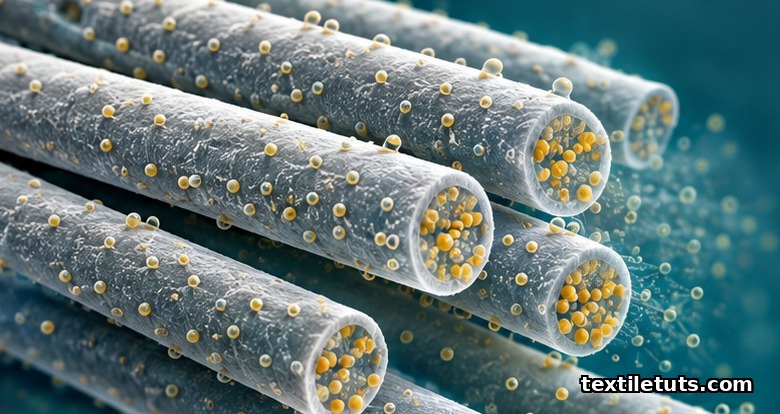
Implantable drug delivery textiles are biodegradable fiber-based scaffolds, typically 10 to 100 micrometers in fiber diameter and loaded with 0.1 to 25 weight percent of a therapeutic agent, that release drugs at controlled rates of 1 to 10 percent per day over 7 to 90 days when placed at a target tissue site.
This local release profile keeps drug concentrations inside the therapeutic window at the diseased tissue while keeping systemic plasma levels 5 to 50 times lower than equivalent oral doses, which is the central reason textile implants are preferred over systemic therapy for many chronic and post-surgical conditions.
The rest of this article walks through the materials, drug-loading methods, release kinetics, biocompatibility standards, and clinical applications that define the field, so a textile engineer or product developer can match a specific drug and indication to the right fiber chemistry and architecture.
How Implantable Drug Delivery Textiles Work
An implantable drug delivery textile is a woven, knitted, braided, electrospun, or nonwoven fabric made from biocompatible and usually biodegradable polymer fibers, with the active drug incorporated inside the fiber matrix, coated onto the fiber surface, or bound to the fiber via a cleavable linker. After implantation, the drug leaves the textile through one or a combination of four mechanisms: diffusion through water-filled pores in the swollen polymer, polymer degradation and erosion, osmotic pumping driven by water uptake, or triggered release in response to a local stimulus such as pH change, temperature, or an enzyme.
The release profile is shaped by three engineering choices: fiber chemistry, fiber geometry, and how the drug is loaded. A poly(lactic-co-glycolic acid) fiber 50 micrometers in diameter that has the drug dissolved inside the polymer matrix will release over 30 to 60 days, while the same polymer as a 5 micrometer electrospun fiber with the drug coated on the surface will release over 3 to 7 days. Choosing a more slowly degrading polymer such as polycaprolactone extends the window to 90 to 180 days. Choosing a hydrogel-forming polymer such as a crosslinked polyethylene oxide shortens it to under 24 hours.
Materials Used in Implantable Drug Delivery Textiles
Three families of polymers dominate the published literature on implantable drug delivery textiles. Each family has a different degradation window and a different regulatory track record.
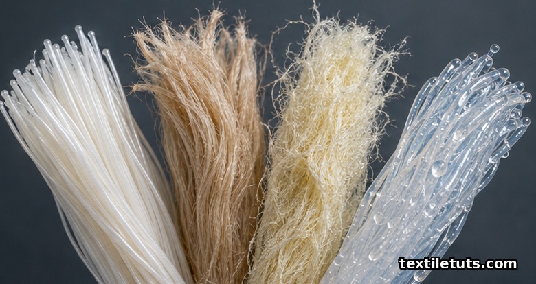
- Synthetic biodegradable polyesters. Poly(lactic acid) (PLA), poly(glycolic acid) (PGA), their copolymer poly(lactic-co-glycolic acid) (PLGA), and polycaprolactone (PCL). PLA, PGA, and PLGA have a long history of FDA-cleared use in resorbable sutures, fixation screws, and tissue scaffolds; PLGA 50/50 loses most of its mass in 1 to 2 months, while high-lactide PLGA and PCL hold their mass for 6 to 24 months.
- Natural polymers. Silk fibroin, collagen, gelatin, chitosan, alginate, and hyaluronic acid. These are degraded by natural enzymes and have structural protein sequences that support cell adhesion, which is useful when the textile is also meant to guide tissue regeneration, not just release drug.
- Hydrogel-forming synthetic polymers. Polyethylene glycol (PEG), polyvinyl alcohol (PVA), and copolymers such as PEG-poly(lactic acid). These absorb 50 to 90 percent water and release drug by diffusion rather than erosion, giving very predictable zero-order release kinetics over hours to a few days.
For non-resorbable implants, such as certain vascular grafts and hernia meshes, polyethylene terephthalate (PET, also called Dacron) and expanded polytetrafluoroethylene (ePTFE) are used as the textile substrate, with the drug held in a separate coating or in a biodegradable fiber blended into the structure.
Drug Loading Methods and How They Set Release
The same drug at the same target dose can produce very different release curves depending on how it is incorporated into the textile. Four loading methods are standard.
- Matrix loading. The drug is dissolved or dispersed in the polymer solution before the fiber is spun. This gives the longest and most reproducible release because the drug has to diffuse through the polymer before it can leave the fiber. Typical drug loadings are 1 to 25 weight percent.
- Surface coating. The finished textile is dipped, sprayed, or padded with a drug solution, often with a polymer binder. Surface loading gives a high initial burst (often 20 to 40 percent of the dose in the first 24 hours) followed by a much slower tail, which is the right shape for antibiotics where you want a strong early kill followed by a maintenance dose.
- Microencapsulation. The drug is first sealed inside microspheres 1 to 50 micrometers across, and the microspheres are then embedded in the textile. Release becomes a two-stage problem: water enters the microsphere, drug diffuses out, then the released drug diffuses through the textile. This is the standard approach when two drugs need to be released on different timelines from the same implant.
- Cleavable linker. The drug is bonded to the fiber polymer through a linker that breaks in response to a specific local signal, such as the lowered pH inside a tumor or the matrix metalloproteinases that are over-expressed at an inflamed site. The implant stays quiescent until it meets the target environment.
Comparison of Common Textile Architectures
The choice of fabric architecture controls the surface area available for drug release, the mechanical properties of the implant, and how easy the surgeon finds it to handle and fix in place.
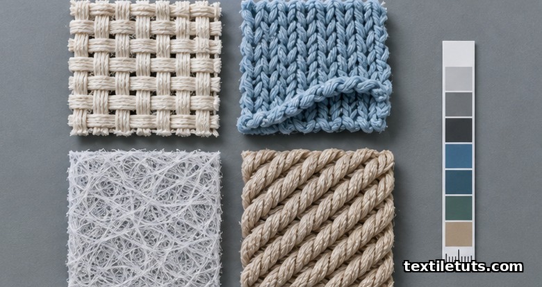
| Property | Woven | Knitted | Electrospun nonwoven | Braided |
|---|---|---|---|---|
| Pore size | 50 to 500 μm | 100 to 1000 μm | 1 to 50 μm | 50 to 300 μm |
| Tensile strength | High | Moderate, high elasticity | Low to moderate | Very high along axis |
| Typical fiber diameter | 10 to 30 μm | 15 to 50 μm | 0.1 to 5 μm | 20 to 100 μm |
| Surface area per gram | 0.1 to 0.5 m²/g | 0.1 to 0.5 m²/g | 10 to 100 m²/g | 0.05 to 0.3 m²/g |
| Typical drug release window | 14 to 90 days | 14 to 90 days | 1 to 30 days | 30 to 180 days |
| Best fit for | Abdominal wall repair, dural patches | Vascular grafts, hernia mesh | Local chemotherapy, wound healing | Tendon and ligament repair, nerve guides |
The very high specific surface area of electrospun mats (often 10 to 100 square meters per gram, two orders of magnitude above woven or knitted fabrics) is the main reason they have become the workhorse for short-duration, high-dose local release in oncology and wound care.
Biocompatibility and Sterilization Standards
Every implantable drug delivery textile must meet the ISO 10993 series for biological evaluation of medical devices before it can enter a clinical trial. ISO 10993-5 covers in vitro cytotoxicity, ISO 10993-6 covers implantation local effects, ISO 10993-10 covers irritation and skin sensitization, and ISO 10993-11 covers systemic toxicity. For drug-device combination products, additional data are required under the United States Food and Drug Administration’s combination product framework, which usually means both the device testing above and the pharmacology, toxicology, and pharmacokinetics of the drug.
Sterilization matters because many common sterilization methods damage the drug or the polymer. Ethylene oxide gas at 37 to 63 degrees Celsius and 40 to 80 percent relative humidity is the most common choice for finished drug-loaded textiles because it runs at temperatures that do not degrade heat-sensitive PLGA or most antibiotics. Gamma irradiation at 25 to 40 kilogray is effective but breaks the chains of some polymers and can change release kinetics by 10 to 30 percent. Steam autoclaving at 121 degrees Celsius is generally incompatible with PLA, PLGA, and most drug-loaded textiles because the heat distorts the polymer above its 55 to 65 degrees Celsius glass transition.
For packaging and shelf life, combination products follow United States Pharmacopeia (USP) General Chapter 1207 for container closure integrity, and accelerated stability data are usually collected at 25 degrees Celsius / 60 percent relative humidity and at 40 degrees Celsius / 75 percent relative humidity to support a 2-year shelf life claim.
Clinical Applications in Practice
Three indications account for most of the published clinical and preclinical work on implantable drug delivery textiles.
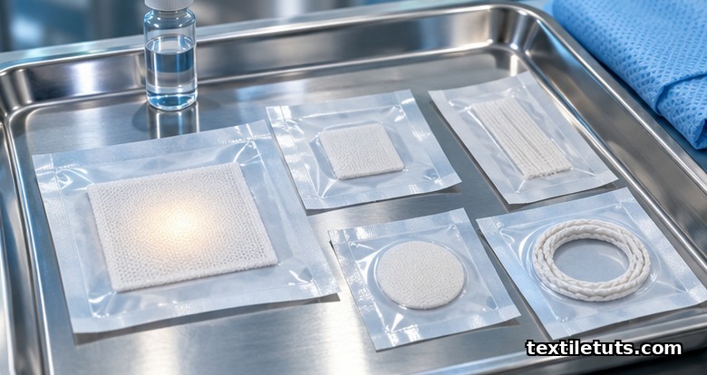
- Post-surgical infection prevention. Triclosan-coated polydioxanone sutures (the Plus suture range) release antiseptic over 14 to 28 days and have been shown in randomized trials to reduce the risk of surgical site infection by about 30 percent compared to uncoated sutures in abdominal closure. The same approach is now applied to hernia meshes and to orthopedic fixation implants.
- Local cancer therapy. Electrospun PLGA mats loaded with paclitaxel, doxorubicin, or cisplatin have been studied for implantation at the tumor resection site in glioblastoma, breast cancer, and head and neck cancer. Local concentrations can be 100 to 1000 times higher than the systemic maximum tolerated dose, which is the central clinical advantage.
- Cardiovascular drug delivery. Drug-eluting vascular grafts and stent grafts are coated or impregnated with heparin, sirolimus, paclitaxel, or other agents to reduce thrombosis and intimal hyperplasia. Heparin release from a polyester graft over 7 to 14 days has been commercially available since the late 1990s.
Other active indications include nerve guidance channels releasing nerve growth factor, dural repair patches releasing anti-fibrotic agents, and tissue engineering scaffolds releasing bone morphogenetic protein-2 for spinal fusion.
Frequently Asked Questions
Q1: How long can an implantable drug delivery textile keep releasing drug inside the body?
Release windows in clinical and preclinical use range from 1 day for a hydrogel-coated mesh to 180 days for a slowly degrading polycaprolactone braided scaffold. The longest published continuous release profiles from a single textile implant are around 12 months, achieved with very high molecular weight polymers and very low water-uptake geometries.
Q2: Are implantable drug delivery textiles safe for MRI?
Most polymeric drug delivery textiles (PLA, PGA, PLGA, PCL, silk, collagen) are MRI-conditional because they do not contain ferromagnetic materials. Any implant that includes metal components, such as a stent graft with metallic stents or a magnetic nanoparticle trigger, must be labeled for MRI per ASTM F2503, and the local heating and image artifact must be characterized.
Q3: What happens to the textile after the drug is finished releasing?
For implants made from biodegradable polymers, the textile loses mass through hydrolysis or enzymatic cleavage and is cleared as carbon dioxide and water (PLA, PGA, PCL) or as small peptides and amino acids (silk, collagen). For non-resorbable substrates such as PET or ePTFE, the textile stays in place permanently, which is acceptable when the implant is also serving a mechanical role such as vascular conduit or hernia reinforcement.
Q4: How is the drug dose for an implantable textile chosen compared to a systemic dose?
The local dose is usually 5 to 50 percent of the total systemic dose, because the drug does not have to be diluted across the whole body. In oncology implants this difference is much larger; published paclitaxel-loaded brain implants release about 1 milligram locally per day, whereas systemic paclitaxel at 100 milligrams per square meter of body surface area would produce severe systemic toxicity at the same local concentration.
Q5: Can a single textile implant release more than one drug on different timelines?
Yes. Microencapsulation, layered coatings, and blended fiber populations are all used. A typical design uses a fast-releasing coating (for example, an antibiotic that finishes in 7 days) layered over a slow-releasing matrix-loaded fiber (for example, an anti-inflammatory that finishes in 60 days), so a single implant covers both the early infection-risk window and the longer healing window.
References
- International Organization for Standardization. ISO 10993-5:2009 Biological evaluation of medical devices, Part 5: Tests for in vitro cytotoxicity. iso.org — the standard test for cell toxicity of medical device materials.
- International Organization for Standardization. ISO 10993-6:2016 Biological evaluation of medical devices, Part 6: Tests for local effects after implantation. iso.org — required for any textile that will be placed in living tissue.
- Langer, R., and Peppas, N.A. (2003). Advances in biomaterials, drug delivery, and bionanotechnology. AIChE Journal, 49(12), 2990-3006. researchgate.net — the foundational review on controlled release from polymer matrices.
- Sill, T.J., and von Recum, H.A. (2008). Electrospinning: Applications in drug and tissue engineering. Biomaterials, 29(13), 1989-2006. sciencedirect.com — covers the use of electrospun nonwovens as drug release matrices.
- Astm International. ASTM F2503-23 Standard Practice for Marking of Medical Devices and Other Items for Safety in the MR Environment. astm.org — the standard that defines MRI-conditional labeling for implants.
- United States Pharmacopeia. General Chapter 1207.1 Package Integrity and Test Methods. usp.org — referenced for combination product container closure.
- International Organization for Standardization. ISO 11135:2014 Sterilization of health care products, Ethylene oxide. iso.org — the standard sterilization method for most drug-loaded textile implants.
This article is the working reference for implantable drug delivery textiles. Editorial by Md. Asad Ullah (Meem), TextileTuts. Sources: ISO 10993-5, ISO 10993-6, ISO 11135, ASTM F2503, USP 1207.1, Langer and Peppas (2003), Sill and von Recum (2008) as cited.


