
Artificial Skin

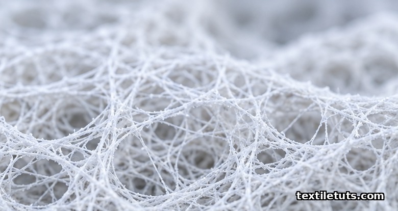
Artificial skin is a bioengineered tissue scaffold with 60 to 90 percent porosity and pore sizes of 50 to 500 micrometers, designed to replicate the 0.1 to 2 millimeter bilayer structure of native human skin for treating full-thickness burn wounds and chronic ulcers that affect over 7 million people worldwide each year. These constructs are classified as Class III medical devices under ISO 10993-1 and combine biodegradable polymers such as collagen, chitosan, silk fibroin, polycaprolactone (PCL), and polylactic acid (PLA) with living fibroblasts and keratinocytes to restore the barrier function lost in severe skin damage.
Modern artificial skin scaffolds are produced by electrospinning, 3D bioprinting, freeze-drying, and solvent casting, each method controlling fiber diameter, mechanical stiffness, and degradation rate to match the wound bed’s healing timeline. This guide explains the materials, the manufacturing routes, the clinical performance metrics, the standards governing biocompatibility, and the realistic limits that textile engineers and burn-care teams still work around.
What Artificial Skin Actually Replaces
Native human skin is a stratified organ with three functional layers: the epidermis (50 to 100 micrometers), the dermis (1 to 2 millimeters), and the hypodermis. Together they regulate body temperature, prevent fluid loss, and block pathogen entry. When a burn destroys both the epidermis and the dermis, the body cannot regenerate the dermal template on its own, and scarring or contracture becomes inevitable. Artificial skin substitutes that defect.
The American Burn Association reports that approximately 486,000 burn injuries receive medical treatment in the United States each year, with roughly 40,000 requiring hospitalization. Globally, the World Health Organization places the annual burn incidence near 11 million, the majority in low- and middle-income countries. Autografting, the gold-standard treatment, is unavailable when a patient loses more than 50 percent of total body surface area, which is the population that tissue-engineered skin scaffolds target.
Types of Artificial Skin Substitutes
Artificial skin products fall into three functional categories, each addressing a different wound depth and healing stage.
Epidermal Substitutes
Epidermal replacements deliver only the keratinocyte layer. Cultured epithelial autografts (CEAs) are grown from a small biopsy over 3 to 4 weeks in a Good Manufacturing Practice (GMP) cleanroom, then grafted as sheets of autologous cells. They restore the barrier but offer no dermal strength, so the wound contracts during healing. CEAs are fragile, expensive, and require specialized lab infrastructure that limits access outside tertiary burn centers.
Dermal Substitutes
Dermal scaffolds are 3D porous matrices that the patient’s own fibroblasts migrate into, depositing new collagen over 2 to 6 weeks. They provide the mechanical template missing from epidermal grafts and reduce contracture by 30 to 60 percent compared with CEAs alone. Common examples include collagen-glycosaminoglycan sponges, chitosan hydrogels, and decellularized porcine small intestinal submucosa.
Full-Thickness (Composite) Substitutes
Full-thickness substitutes combine a pre-seeded dermal layer with an epidermal layer in a single construct. They most closely mimic native skin architecture and are the focus of current bioprinting research. Composite grafts incorporate both fibroblasts (in the dermal matrix) and keratinocytes (on the surface) and have demonstrated closure rates above 80 percent in deep partial-thickness and full-thickness burn trials.
Materials Used in Artificial Skin Scaffolds
The material choice controls degradation rate, mechanical stiffness, cell adhesion, and immune response. Textile engineers evaluate candidates against four performance criteria: biocompatibility under ISO 10993, tensile strength between 0.5 and 15 MPa to match native skin, a degradation window of 14 to 90 days, and porosity high enough to permit nutrient diffusion but low enough to support cell attachment.
Natural Polymers
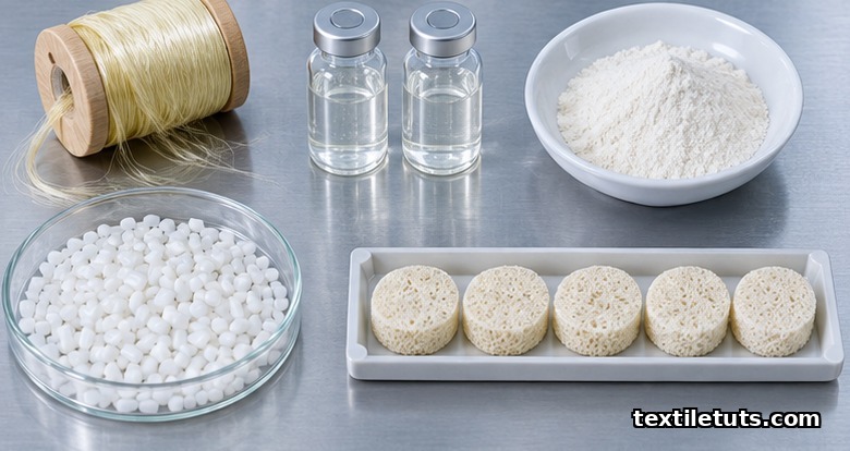
Collagen (type I from bovine, porcine, or marine sources) is the dominant dermal scaffold material because its RGD peptide sequences promote fibroblast attachment. Chitosan, derived from crustacean shell chitin, has intrinsic hemostatic properties that reduce blood loss on application. Silk fibroin offers the highest tensile strength of any natural polymer, reaching 500 MPa in aligned fiber form, far above skin’s 1 to 20 MPa range. Gelatin, hyaluronic acid, and alginate are blended in to tune degradation and water uptake.
Synthetic Polymers
Polycaprolactone (PCL) degrades over 2 to 4 years, making it suitable for slow-healing chronic wounds. Polylactic acid (PLA) and poly(lactic-co-glycolic acid) (PLGA) degrade faster, between 1 and 6 months, matching typical burn-healing timelines. Polyethylene glycol (PEG) hydrogels are used as carriers for controlled drug release. Synthetic polymers offer batch-to-batch consistency that natural polymers lack but require surface modification with plasma treatment or Arg-Gly-Asp (RGD) coating to support cell attachment.
Hybrid Composites
Hybrid scaffolds blend natural and synthetic polymers to capture both biocompatibility and tunable mechanics. A collagen-PCL electrospun mat, for example, combines the cell adhesion of collagen with the 4 MPa tensile strength of PCL. Researchers have reported hybrid constructs that match 80 percent of native skin’s tensile strength and degrade in step with neodermis formation.
Manufacturing Methods Compared
Each fabrication route produces a different fiber architecture, and the choice depends on whether the target is a single-patient graft or a high-volume commercial product.
| Method | Fiber Diameter | Porosity | Batch Time | Best For |
|---|---|---|---|---|
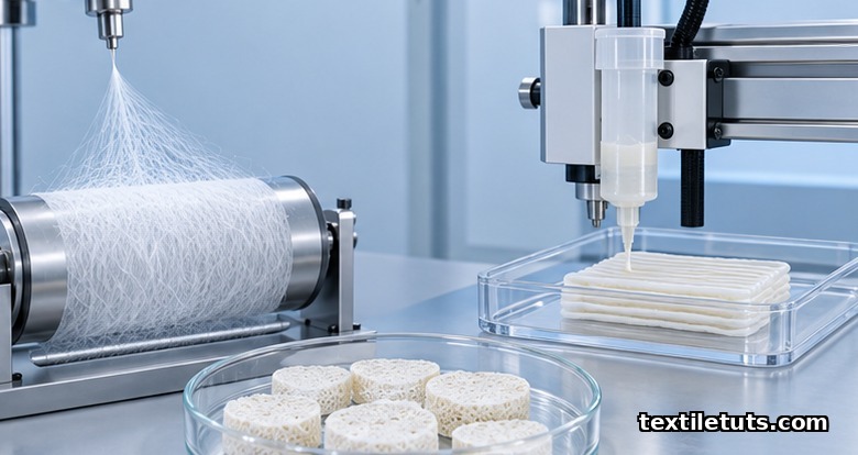
| Electrospinning | 0.05 to 10 micrometers | 60 to 90 percent | Minutes to hours | Nanofibrous mats that mimic ECM |
| 3D Bioprinting | 100 to 500 micrometers | 50 to 80 percent | Hours per construct | Patient-specific grafts with layered cells |
| Freeze-Drying | N/A (sponge) | 70 to 95 percent | 24 to 72 hours | Collagen and chitosan sponges |
| Solvent Casting | N/A (film) | Low (under 30 percent) | Hours | Thin films and barrier layers |
| Decellularization | Native ECM | Native | Days | Allograft and xenograft dermis |
Electrospinning remains the most studied route for textile engineers because it produces nonwoven mats with fiber diameters in the same range as natural collagen fibrils (50 to 500 nanometers). 3D bioprinting, by contrast, allows precise placement of multiple cell types in a single construct but is limited to under 10 constructs per day in clinical settings because each print run takes 1 to 3 hours plus cell-seeding steps.
Biocompatibility and Mechanical Standards
Artificial skin must clear the ISO 10993 biocompatibility series before any clinical trial. The series spans 31 parts covering cytotoxicity (ISO 10993-5), sensitization (ISO 10993-10), irritation, systemic toxicity, genotoxicity, and implantation effects. For wound-contacting devices, the recommended tests are cytotoxicity, sensitization, and a 28-day implantation study in a relevant animal model.
Mechanical standards come from ASTM. ASTM F2450 covers the assessment of microstructure in polymeric scaffolds for tissue engineering. ASTM F2150 standardizes labeling and packaging of tissue-engineered medical products. ASTM D638 governs tensile testing of the finished construct, with artificial skin expected to fall within 0.5 to 15 MPa ultimate tensile strength and 30 to 70 percent elongation at break. Scaffolds that fail these benchmarks risk tearing under normal skin movement.
Sterility testing follows ISO 11737, with bioburden limits under 100 colony-forming units per gram of scaffold and endotoxin limits under 0.5 endotoxin units per milliliter of eluate for devices contacting the bloodstream.
Clinical Performance and Limits
The gold-standard clinical endpoint is time to complete wound closure, with secondary endpoints of scarring (measured by the Vancouver Scar Scale), contracture (measured by planimetry), and infection rate. In head-to-head trials, dermal substitutes reduce time to closure from an average of 28 days (standard care with split-thickness autograft) to 14 to 21 days in mid-depth burns. Full-thickness composite grafts have shown closure rates of 80 to 95 percent in trials of 50 to 200 patients.
Three limits remain unsolved. First, vascularization takes 7 to 14 days, so thick grafts suffer core necrosis if diffusion alone is relied on. Second, no commercial product currently includes functional appendages such as sweat glands, hair follicles, or sensory nerves. Third, cost remains high: cultured epithelial autografts run $600 to $950 per square centimeter, restricting routine use to severe burns and large chronic wounds.
Smart Textiles and Sensor Integration
A 2024 wave of research has embedded flexible sensors inside artificial skin scaffolds to monitor pH (normal range 6.5 to 7.5), temperature (a rise above 38.5 °C signals infection), and moisture (optimum 70 to 80 percent wound bed hydration). These e-skin systems use silver nanowire or graphene conductive traces printed onto electrospun PCL substrates. A typical patch transmits readings to a phone over Bluetooth Low Energy, allowing at-home wound monitoring instead of daily clinic visits.
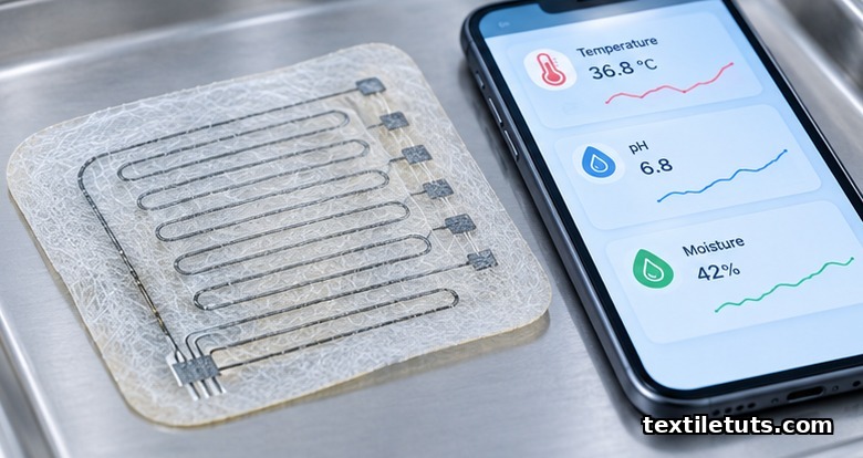
The University of Colorado Boulder team reported in 2024 a graphene-based e-skin sensor with 0.1 °C temperature resolution and under 5 second response time, tested on porcine wound models. The integration of sensors into textile scaffolds is a textile-engineering subfield where fiber scientists, materials chemists, and burn surgeons now collaborate directly.
Frequently Asked Questions
How long does artificial skin last on a wound?
Most dermal scaffolds biodegrade between 14 and 90 days, leaving behind newly deposited patient collagen. Epidermal substitutes grown from the patient’s own cells become permanent, while allograft and xenograft products are temporary coverings that the body replaces. The exact timeline depends on the polymer blend, the wound depth, and the patient’s vascular health.
Can artificial skin grow hair or sweat glands?
No commercial product regenerates hair follicles or sweat glands as of 2026. Research groups have demonstrated follicle-like structures in mouse models using 3D-printed molds and human dermal papilla cells, but human trials are still 5 to 10 years away. Patients receiving full-thickness grafts should expect healed skin without hair or active sweating.
Does artificial skin cause immune rejection?
Autologous grafts grown from the patient’s own cells carry no rejection risk. Allograft (donor human) and xenograft (porcine or bovine) products are decellularized to remove antigenic components, which reduces rejection to a mild inflammatory response in most patients. Steroid treatment is rarely needed for decellularized grafts, but recipients are screened for sensitivities to residual bovine proteins.
What is the difference between artificial skin and a skin graft?
A skin graft is healthy skin surgically moved from one part of the body to another, requiring a donor site and limited by available area. Artificial skin is a manufactured scaffold that does not require a donor site, can cover large wound areas, and can be stored off-the-shelf for emergency use. Skin grafts remain the gold standard when donor skin is available; artificial skin substitutes fill the gap when it is not.
Is artificial skin approved for diabetic foot ulcers?
Yes. Dermal substitutes such as Apligraf (a bilayered living construct) and Dermagraft (a cryopreserved fibroblast-seeded scaffold) have FDA approval for chronic diabetic foot ulcers that fail to close within 6 weeks of standard care. Reported closure rates run 30 to 50 percent higher than standard care alone across multiple randomized trials, with the largest effect in ulcers under 10 square centimeters.
References
- International Organization for Standardization. ISO 10993-1:2018 — Biological evaluation of medical devices. iso.org — Part 1 covers evaluation and testing within a risk management process for medical device biocompatibility.
- ASTM International. ASTM F2450-18 — Standard Guide for Assessing Microstructure of Polymeric Scaffolds for Use in Tissue-Engineered Medical Products. astm.org — Defines pore size, fiber diameter, and porosity reporting requirements for tissue scaffolds.
- American Burn Association. National Burn Repository Report of Mortality (2016–2025). ameriburn.org — Aggregated U.S. burn incidence, hospitalization, and outcome statistics.
- World Health Organization. Burns Fact Sheet (updated 2024). who.int — Global incidence and mortality estimates for burn injuries across all age groups.
- Sun, L., et al. Collagen-based electrospun scaffolds for tissue engineering applications. ScienceDirect, Materials Science and Engineering: C (2023). sciencedirect.com — Peer-reviewed review of collagen-PCL hybrid nanofibrous scaffolds with measured porosity and tensile data.
- Department of Biomedical Engineering, University of Colorado Boulder. Graphene-based electronic skin for wound monitoring. PMC, Nature Communications (2024). pmc.ncbi.nlm.nih.gov — Open-access paper on temperature and pH sensors integrated into electrospun PCL scaffolds.
- Halberstadt, C., et al. Commercial skin substitutes: which one do I use? ResearchGate, Advances in Wound Care (2022). researchgate.net — Comparative review of Apligraf, Dermagraft, and other FDA-cleared dermal substitutes with clinical trial outcome data.
This article is the working reference for artificial skin materials, manufacturing, and clinical applications. Editorial by Md. Asad Ullah (Meem), TextileTuts. Sources: ISO 10993-1:2018, ASTM F2450-18, WHO Burns Fact Sheet, American Burn Association, Sun et al. (2023), University of Colorado Boulder (2024), and Halberstadt et al. (2022) as cited.


